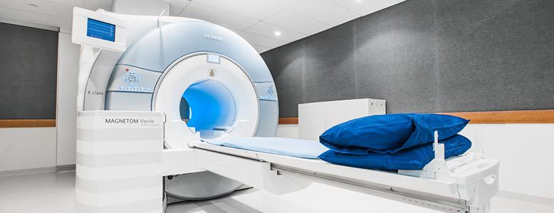
There is increasing evidence that multiparametric MRI of the prostate (mpMRI) has an important role, not only in the staging of prostate cancer, but also in the diagnosis.
It adds significantly to our current screening tools of Prostate Specific Antigen blood test and Digital Rectal Examination and aids in the selection of patients who require a prostate biopsy. If a significant lesion is identified on the mpMRI, the MRI can then be used to accurately target the lesion in the form of a MRI guided prostate biopsy.
Magnetic Resonance Imaging (MRI) is a type of scan that provides detailed images of soft tissue such as your prostate.
MRI produces different information when compared to other examinations such as ultrasounds; CT scans or x-rays and in particular provides information concerning soft tissue. The MRI machine uses a strong magnetic field and radio waves to examine your prostate and does not use x-rays; it is safe and painless.

MRI techniques have improved in recent years and MRIs’ utility in aiding diagnosis, tumour localisation and staging, assessment of aggressiveness, treatment planning and follow up in patients with prostate cancer is well established. MRI improves visualisation of the prostate, its substructure, surround tissues and most importantly focal lesions or cancer. MRI is a versatile and promising technique and appears to be the best available imaging technique for the prostate.


